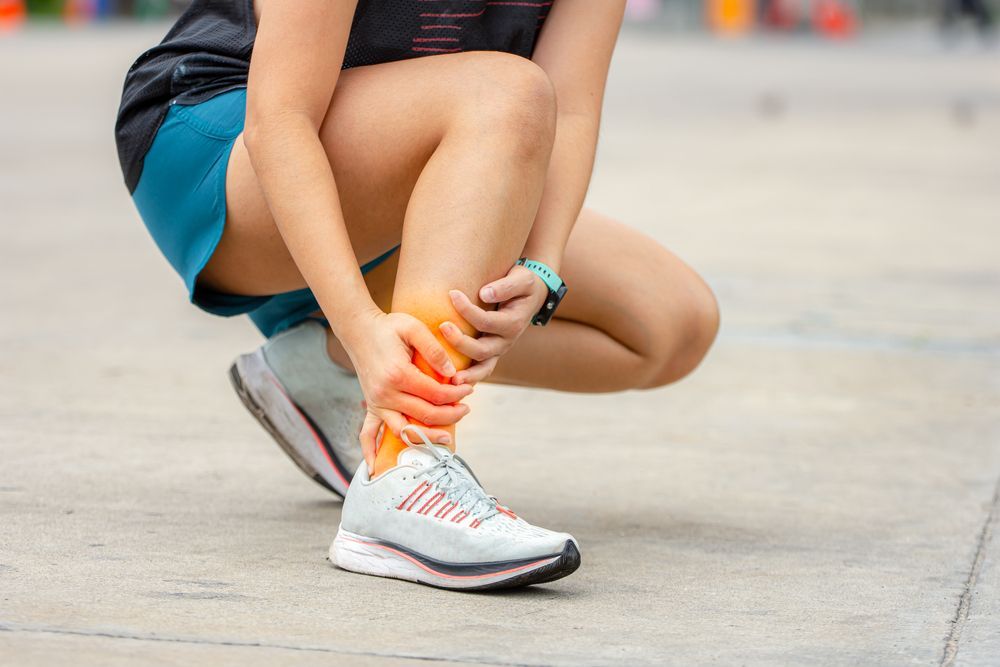

Achilles Tendinopathy
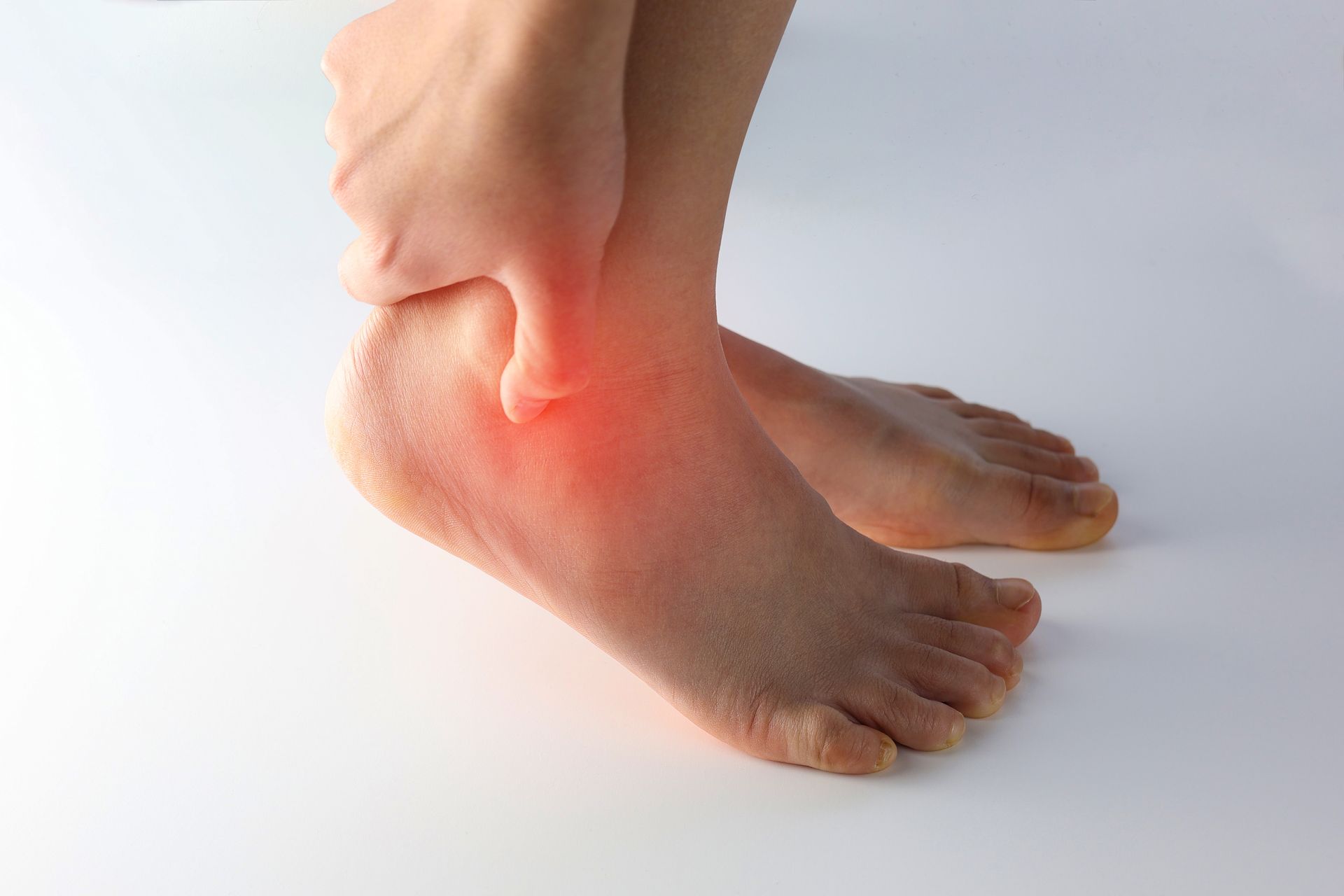
Achilles Tendinopathy-
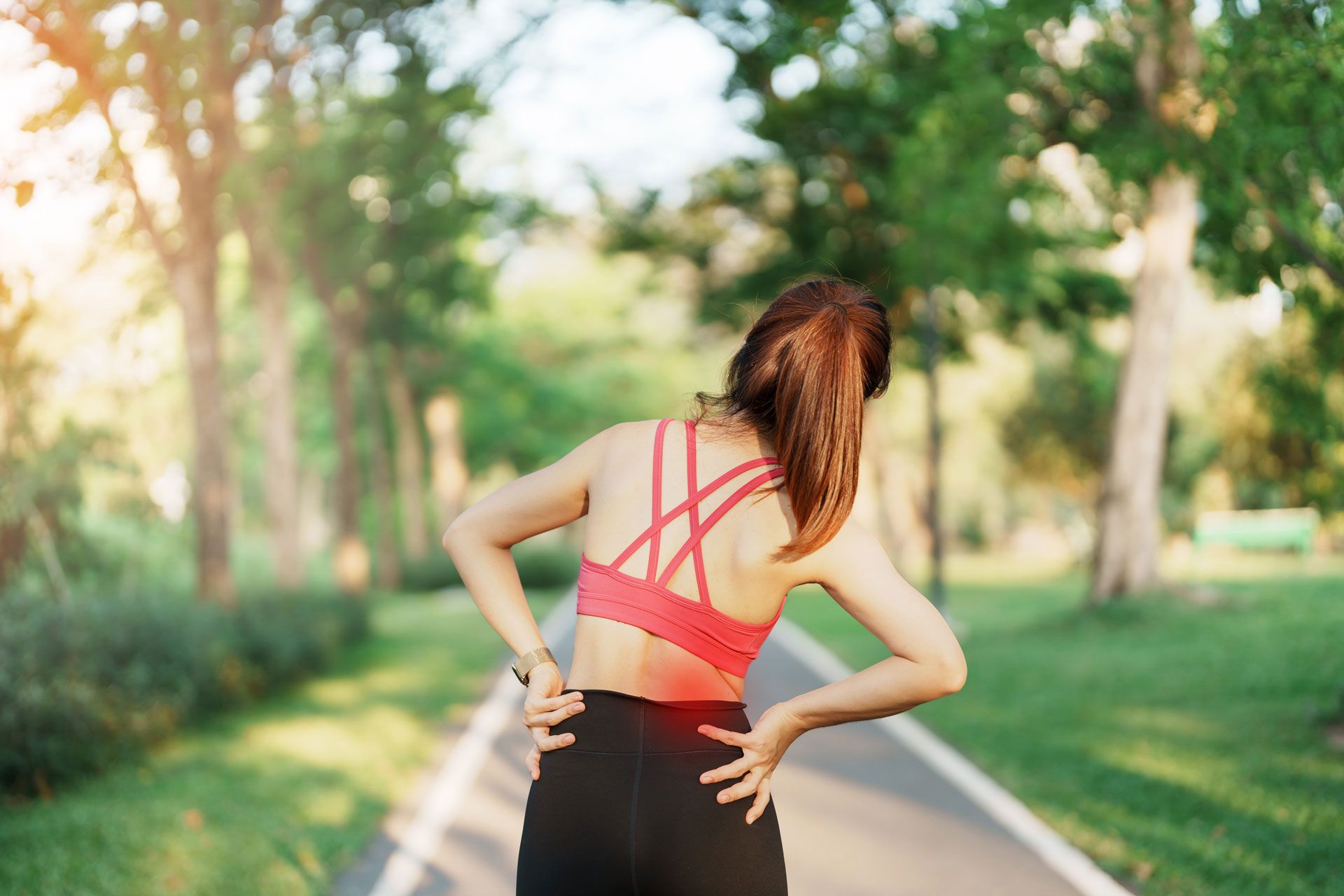
is an overuse injury to the Achilles tendon which is common in sports that involve running and jumping. However, in general practice 65% of the cases are not sports related. Recovery can take up to a year and recurrent rate is high, especially if the rehab and return to activities is rushed. The key to successful outcome hinges on early intervention, comprehensive treatment and appropriate activity modification.
Exercise rehab is the key treatment for Achilles tendinopathy and is divided into four stages: symptom management, recovery, rebuilding, and return to activity/sport. The ultimate goal is to improve tendon strength, tensile capacity and endurance with a return to full activities.
Stage One: Symptom Management
Modify activity – to stop overloading and cumulative micro trauma to the tendon. Having pain after activity that lasts longer than 48 hours indicates that activity needs to be reduced. Cardio vascular activities are essential in the healing process therefore off-loading activities such as cycling, water aerobics, and swimming will speed up the heeling process.
Strengthening – standing heel raises on a flat surface, if its painful place a lift under the heel. 3x15-20, 2 second pause at the top and bottom. It is important the heel raises are done in the pain-free range of motion. Immobilization and complete rest can actually delay recovery and is not recommended.
Stage Two: Recovery
The goal here is to strengthen the tendon and improve its tensile strength. Steadily increase the load on the Achilles by performing heel raises off a step and allowing the heel to lower below the edge, 3 X 15-20 reps. Slowly go as deep as you can without pain and hold for 2 secs. Progress to single leg eccentrics by going up on both feet, remove one foot and slowly lower on the other leg. Again, build to 3 × 15-20 reps with no increase in symptoms. Be sure to maintain cardio vascular fitness levels.
Stage Three: Rebuilding
The objective in the rebuilding phase is to steadily increase the strength and endurance of the entire calf, hamstring and glute musculature to prepare for full activities.
Increasing load can simply be done by having the patient wear a weighted backpack while doing the same heel raises as in the recovery stage. Smith machine, standing calf raises, seated calf raises, and leg press machines can all be used.
Eccentric loading. jumping and hoping are essential in this stage. Start with 30 jumps with a soft landing one time per day. Build to 5 sets then progress to jumping rope at 30 second intervals and finally single leg hops, 3 X 15. Continue the heel raise exercises while adding in the plyometrics.
Stage Four: Return to Activity/Sport
Return to full activities requires strength and tissue compliance. Minimal symptoms, with intermittent stiffness in the morning, is acceptable and the level of activity can be maintained. Continue the weighted single leg exercises on the edge of a step, (concentric and eccentric), jumping and hoping activities. If symptoms escalate and last more than 24 hours, take a day off and peel back the exercises to a level where there is no pain after 24 hours. Then slowly increase.
Achilles tendinopathy can be slowly resolving, and you can discuss with the doctor the goals and objectives of each sage of care to avoid reinjury. Especially when you "feel pretty good" and want to ramp up activity. Be sure to maintain cardiovascular fitness, core, frontal plane stability and total body conditioning by cross training in all stages of care.
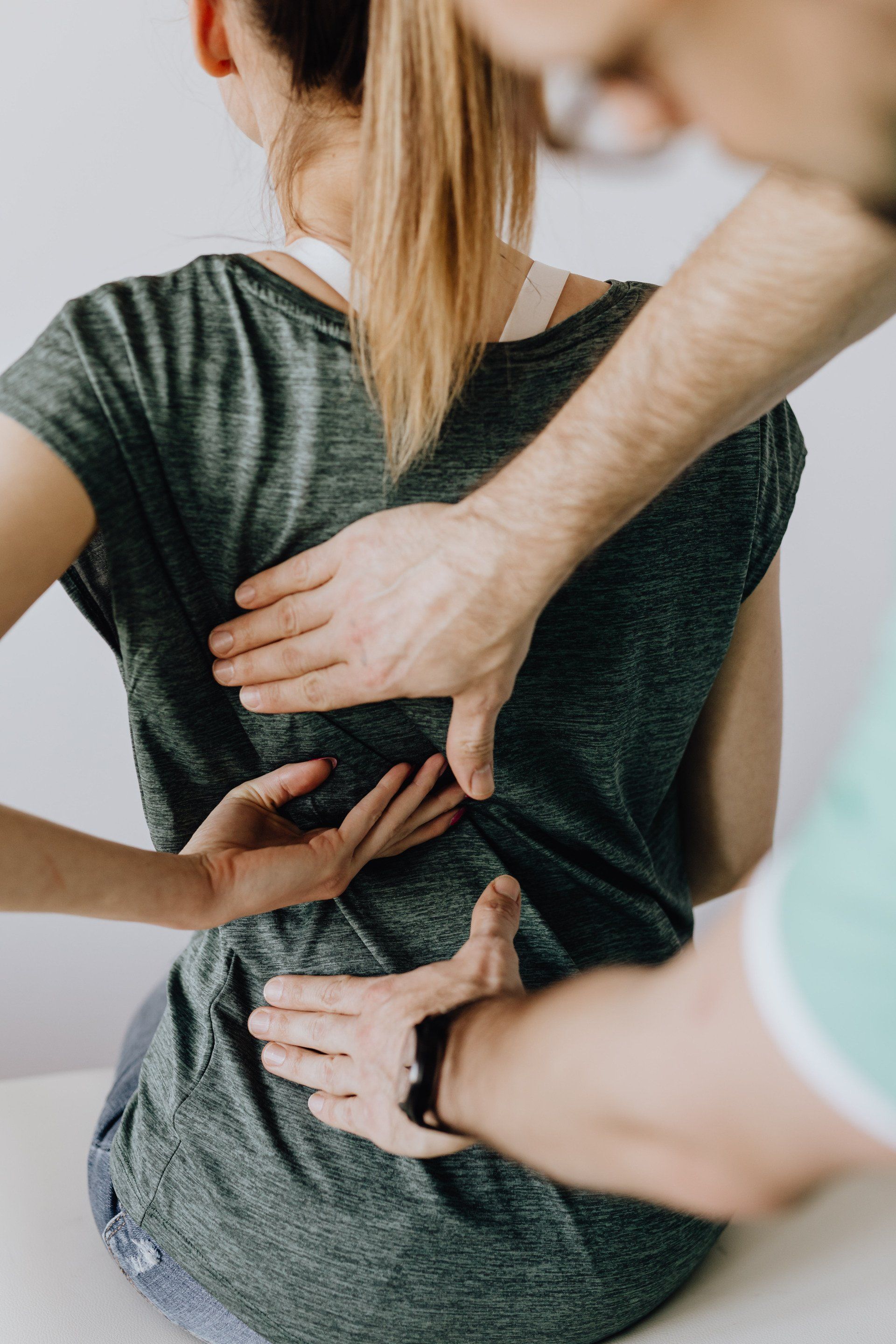
Chiropractic rehabilitation, however, is more than exercises.
As a chiropractor, I make sure to check and adjust the feet, ankles, knees and hips as needed. We perform soft tissue work both locally and along the entire fascial chain. Adding nutraceuticals such a collagen, vitamin C and branched chain amino acids. Finally, incorporating modalities to increase blood perfusion to the region such as FAKTR, EPAT (radial shockwave therapy), and Active Release Technique will accelerate the response to treatment.